Label the Image to Test Your Understanding of Antibody Structure
It consists of four polypeptide chains two heavy H chains and two light L chains. Select characteristics of the lymphatic system to test your understanding of its structure and major functions.

Antibodies 101 Introduction To Antibodies
We review their content and use your feedback to keep the quality high.

. Aspen Independent Dement - T-cell line dance Antigen Antibodies Plasma cells T cytotoxic Activated T cel Eosinophils АРС B-cell line. Label the image to test your understanding of antibody structure Carbohydrates Hirgo regions Ode bords 11111 Complement Antigen binding the 19 of 20 Next. While one part of the antibody the antigen binding fragment Fab recognizes the antigen the other part of the antibody known as the crystallizable fragment Fc interacts with other elements of the immune system such as phagocytes or components of the complement pathway to.
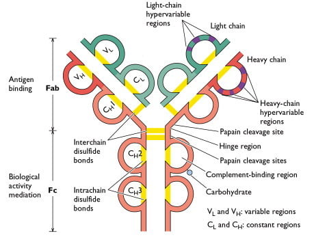
Antibodies are our molecular watchdogs waiting and watching for viruses bacteria and other unwelcome visitors. Light chains consist of the CL and Vκ or Vλ elements. In this article we will discuss about the structure of antibody with the help of suitable diagram.
I hope by the above labelling. This quiz and corresponding worksheet will test you on your knowledge of antibody structure. In humans there are five chemically and physically distinct classes of antibodies IgG IgA IgM IgD IgE.
Top to bottom Antigen-Independent Development. Drag the images to their corresponding class to review the structure and function of antibodies. The region holding arms and stem of antibody is termed as hinge.
Label the image to test your understanding of lymphocyte development and function within the third line of defense. Label the image to test your understanding of antibody structure Carbohydrates Angeling de Oo I Next. Immunoglobulin G IgG the most abundant type of antibody is found in all body fluids and protects against bacterial and.
Biology questions and answers. When they find an unfamiliar foreign object they bind tightly to its surface. We review their content and use your feedback to keep the quality high.
Each H chain is comprised of the constant region Cα1 Cα2 Cα3 hinge region and the Variable V region. Antibodies circulate in the blood scrutinizing every object that they touch. Such a reaction can be viewed only under the microscope by a labeled antibody.
View the full answer. There is a printable worksheet available for download here so you can take the quiz with pen and paper. The HV regions of a Fab representing both light and heavy chains are highlighted in purple.
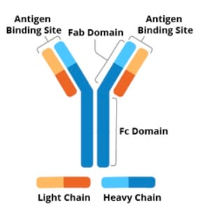
The aim of this part of the chapter is to explain how this structure is formed and how it allows antibody molecules to. Drag the images to their corresponding class to review the structure and function of antibodies. They have a Y shaped structure.
Light chain heavy chain antigen binding site Fc Fab domains hinge region disulfide bonds and variable region. The four polypeptide chains are held together by disulfide bonds to form a Y shaped structure. IgG Most prevalent antibody in circulation IgA Dimer that is a significant component of mucus and secretions Antibody made at the beginning of initial response to antigen IgM Main function is to serve as antigen receptor.
Paradigm of antibody antigen recognition is that the three-dimensional structure formed by the six CDRs recognizes and binds a complementary surface epitope on the antigen. Match the correct statement with the antibody type to test your understanding of structure and functions of antibodies. A label can be an enzyme a fluorophore or colloidal gold.
IgA antibody structure and function. Label the image to test your understanding of lymphocyte development and function within the third line of defense. Certain antibodies get attached to specific antigens.
Although CDR loops are hypervariable and confer binding speci city to the antibody it is not necessary that all six CDR loops interact with a given antigen. The antigen is green. Label the image to test your understanding of antibody structure.
This reaction forms the basis of identification tests. Who are the experts. Match the statements to the term they describe to test your understanding of the structure of an antibody molecule.
Lymphocyte Stock Images by urfingus 6 132 The structure of a typical antibody moleculeAntibodies and amino acids Stock Photos by creativepic 0 0 Immunoglobulin molecule Picture by LeonidAndronov 2 52 The structure of a typical antibody moleculeAntibodies and amino acids Stock Photography by creativepic 0 0 Human papillomavirus HPV 16. Label the image to test your understanding of how antigens are presented to T cells. Antibodies or immunoglobulins are glycoproteins that bind antigens with high specificity and affinity.
Experts are tested by Chegg as specialists in their subject area. They are also known as immunoglobulins Igs. Antibody molecules are roughly Y-shaped molecules consisting of three equal-sized portions loosely connected by a flexible tether.
Experts are tested by Chegg as specialists in their subject area. Label this figure to demonstrate your understanding of T-cell activation. Label the images to test your understanding of the structure and function of both the lymphatic and mononuclear phagocyte systems.
Immunoglobulin A IgA antibodies consist of heavy H and light L chains. Choose the statement that best describes the primary action of B cells. The other images in this section are derived from this structure.
This image represents the structure of an antibodys variable region Fab complexed with an antigen in this case hen egg white lysozyme. - Form specialized plasma cells that produce antibodies - Mature in the thymus. Antibodies belong to a group referred to as gamma globulins and are called immuno globulins.
The chemical substance produced by the body against the antigen is called antibody. In the case of viruses like rhinovirus or poliovirus a coating of bound. Draw the fundamental structure of an immunoglobulin.
Three schematic representations of antibody structure which has been determined by X-ray crystallography are shown in Fig. An antibody binds to an antigen to start an immune response. The main function of IgA is to bind antigens on microbes before.
In simplistic terms antibodies perform two main functions in different regions of their structure. June 24 2018 by Sagar Aryal. Also learn about the production of monoclonal antibodies.
This is an online quiz called Antibody Structure. Youll need to know about concepts such as light and heavy chains and. Label the parts and be sure to label.

Sketch And Label Structure Of Antibody


0 Response to "Label the Image to Test Your Understanding of Antibody Structure"
Post a Comment